
This post is also available in Dutch.
What is PET?
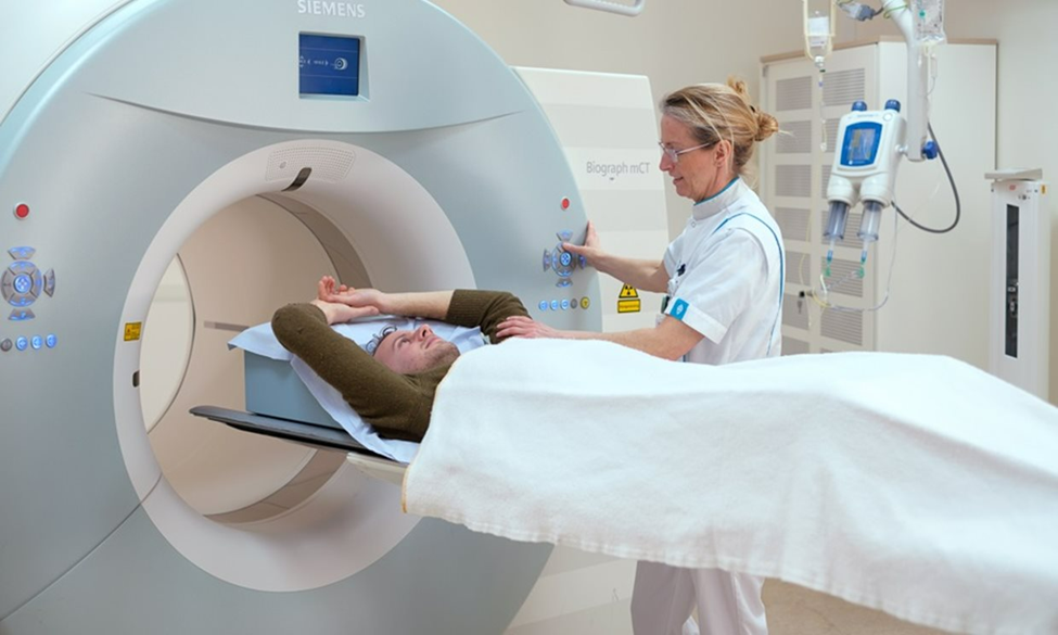
Positron emission tomography (PET) is a common imaging technique that visualizes the metabolism of a radioactive substance (or tracer). The tracer is injected into a blood vessel right before a PET scan. The scanning machine (see image below) detects the subatomic particles emitted by the tracer and later absorbed by the organ of interest (which is often the brain, at Donders Institute). PET imaging can be used for various research purposes, depending on the tracer. For example, fluorodeoxyglucose (FDG) is one of the most commonly used tracers as it indicates glucose (blood sugar) consumption. Therefore, the FDG tracer can be used to visualize energy consumption in different brain areas. Just like most radiotracers, FDG is similar to naturally occurring glucose so your body treats it in a similar way. The radiotracer will usually pass out of your body naturally within a few hours.
PET used in psychology and neuroscience
Previous posts by Donders Wonders introduced fMRI, MEG and EEG as important research methods used by psychologists and neuroscientists. While MEG and EEG are good at finding out when brain activity happens, MRI is good at locating where brain activity happens. Likewise, PET imaging of the brain can also be used to study cognition and behavior:
- For example, a previous study used PET scanning as participants performed various motor and sensory tasks. Specific brain regions that became active during tasks were also correlated with other brain imaging methods, including EEG and fMRI.
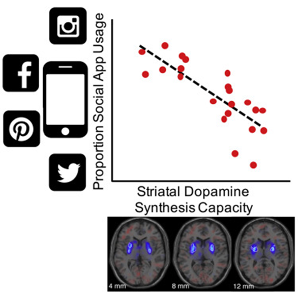
- Additionally, a recent study at the Donders Institute used PET imaging to measure how much dopamine our brain can produce (dopamine is an important brain chemical mainly involved in motivation and reward). However, the capacity of how much dopamine can be synthesized differs largely across people. Researchers found that these individual differences in dopamine synthesis capacity were related to a wide variety of cognitive functions, including motivation, cognitive effort (i.e., effort you make mentally, such as staying focused) , and even smartphone social activity.
Doctors use PET to identify potential disease
Given the high cost of PET in time and money as well as the high demand for medical use, most PET machines are located in hospitals rather than in research institutes. Doctors often use PET to make clinical judgments. For example, as mentioned earlier, PET is used to visualize energy consumption in different brain areas via the FDG tracer. Because most infections and inflammatory cells have higher sugar metabolism, doctors can use this method to identify potential medical conditions in various organs, such as the brain, heart, lung, and so on. Therefore, PET imaging is of great value to clinical trials as well as to scientific researchers.
Curious to see how a PET scan works? Then watch this video.
Author: Ping Chen
Buddy: Christienne Damatac
Editor: Christienne Damatac
Translation: Wessel Hieselaar
Editor translation: Marlijn ter Bekke
Cover image credit to Erik Karits from Unsplash