
This post is also available in Dutch.
Electric messages travel through your brain
As you may already know, the brain consists of lots of brain cells, also known as, neurons. These neurons transmit electrical and chemical signals to each other. If a neuron is stimulated strongly enough, for example by something happening in our environment, an action potential is created. When this happens, gates on the cell wall of the neuron open and close, and particles with a positive or negative electrical charge flow in and out of the cell. This influx and outflow of these particles change an electrical charge in the neuron that travels super-fast through the neuron’s long tail, the axon. At the end of the cell, the neuron sends a signal to surrounding neurons. This is how a message travels through your brain.
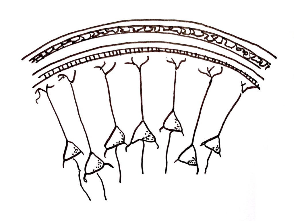
Many neurons summing together create a measurable signal: the current dipole
But how can we measure this message? First, a neuron never “fires” on its own, but with many others at once. We can think of each neuron as a tiny battery. If a group of brain cells fire at about the same time and the current flows in about the same direction, then the electrical signals of all those little batteries add up to become one big battery. We can then measure the sum. But because action potentials are super-fast, neurons hardly fire exactly at the same time. Instead, what we measure is not so much the action potential itself, but more the aftermath. The charged particles that entered the brain cell and flowed through the neuron at lightning speed now flow back outside the cell, slowly enough that it happens at the same time as neighbouring neurons. In the outer shell of the brain, the cortex, there are many pyramidal cells all oriented in the same direction – Perfect for measuring such summation! We call this summation the current dipole.
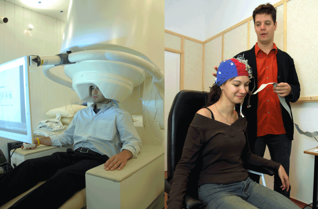
An electric current in your brain can be measured with EEG or MEG
The first method of measuring the electrical current in your brain is with electrodes that we put on your head that pick up the difference in electrical charge. This is called an electroencephalogram, or EEG. The second method is a magnetoencephalogram, MEG for short. With MEG, we measure the magnetic field that is generated by the electric current. The MEG equipment is super-sensitive and is located in a special chamber with thick walls to keep out other magnetic fields. This makes MEG less flexible than EEG; however MEG does give a better measurement because magnetic fields can easily pass through the skull and are therefore more measurable.
But neither method is particularly precise in determining exactly where in the brain the activity is occurring. For that, <fMRI link to blog> is more suitable. The beauty of EEG and MEG is that they are very accurate in determining when the brain is active, which fMRI is not so good at. So which method we should use depends on the research question.
Would you like to participate in (EEG/MEG) research? Sign up via SONA.
Author: Floortje Bouwkamp
Buddy: Judith Scholing
Editor & Translation: Brittany van Beek
Editor translation: Rebecca Calcott