This post is also available in Dutch.
This is part of a Donders Wonders series where we look at different tools for studying the brain. In this part, we will look at how we use magnetic resonance imaging (MRI) to investigate the anatomical structure, communication pathways, and chemistry of the brain.
Anatomical structure – Structural MRI
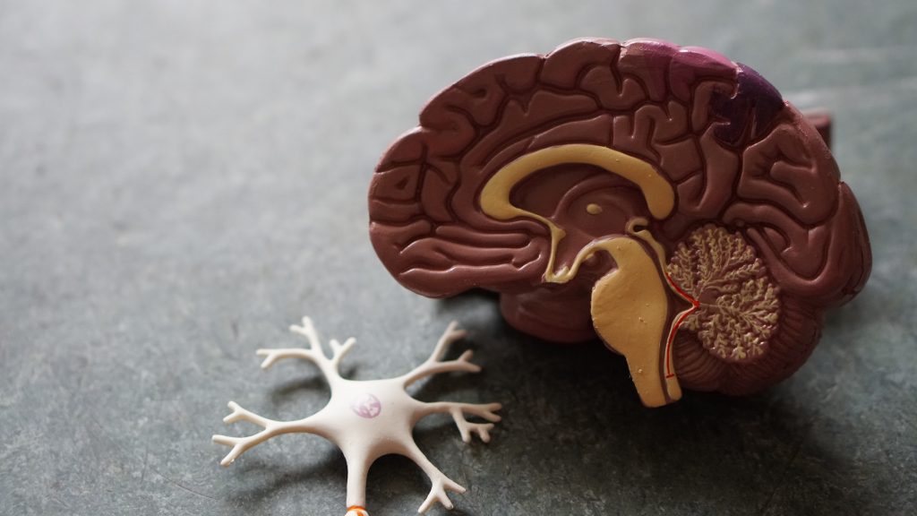
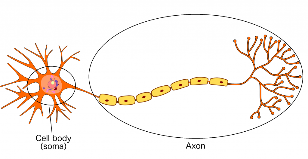
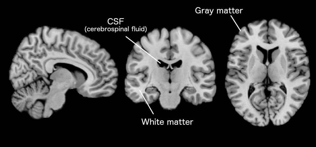
To understand how we get an image of brain anatomy, we need to understand the brain cell, or neuron. It mainly consists of the cell body (soma) and what could be considered the ‘wire’ or ‘cable’ of the neuron, called the axon. The axon connects the neuron to other neurons in the brain. There are two main kinds of tissue in the brain: gray and white matter. Gray matter mainly consists of the somas of neurons and white matter mainly consists of axons. There is also cerebrospinal fluid in and around the brain.
When we make a structural MRI scan, we measure the reaction of water molecules in these tissues to the magnetic field. Because the tissues contain different amounts of water, we can see the differences between them in our images.
Communication pathways – Diffusion imaging
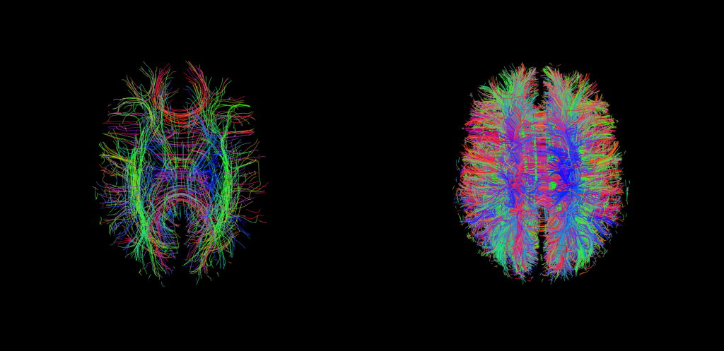
Anatomical scans can measure white matter volume but they neither measure separate axon bundles nor tell us in what direction the axons are going. For this, we can use diffusion-weighted imaging. Water molecules naturally move, or diffuse, more along the length of axon bundles (rather than across). Comparatively, in gray matter and cerebrospinal fluid, water movement direction is more random. Diffusion-weighted imaging measures the movement of water molecules in the brain to estimate the location, diameter and orientation of axon fibres, thus mapping physical communication pathways between brain regions. In the images below, you can see different colors representing different identified pathways.
Chemistry – Magnetic Resonance Spectroscopy
The activity of neurons in the brain depends not only on things like electrical signals and blood flow but also on different chemicals. These chemicals, also known as metabolites, can be measured using MR spectroscopy, or MRS. This is a different imaging method compared to the others because we cannot measure the whole brain at once and, instead, choose a region of interest. MRS then lets us estimate the concentrations of the metabolites in our selected area of interest. Specifically, different metabolites give different signals as a reaction to the magnetic field and we measure this as the chemical shift of each metabolite in our signal. These are metabolites that we cannot measure using positron emission topography, or PET (described in a different part of this series). Instead of giving us colorful pictures of the brain, MRS provides a graph of peaks in the chemical shifts of the metabolites we measure, and we get an image showing us where we measured this in the brain.

Author: Viola Hollestein
Buddy: Rebecca Calcott
Editor: Christienne Damatac
Translation: Floortje Bouwkamp
Editor translation: Felix Klaassen