This post is also available in Dutch .
This blog is part of this year’s summer series called Brain basics, in which we dive into the general functions and development of our brains.
Neurons
Neurons communicate with each other by sending and receiving neurotransmitters, which are chemicals that carry information. Inside of a neuron, the information is passed on through electrical signals. The basic structure of a neuron consists of the dendrite, which receives signals; the soma, which is the main body of the cell; and the axon, which sends signals. There are different kinds of neurons with different shapes and goals, but they all have this structure.

Neurons are connected to each other between one’s axon (sender) and another’s dendrite (receiver). They connect, but never touch. The gap between the neurons is called the synapse. Here, neurotransmitters are released, transmitting the signal between neurons. These signals affect the receiving neuron’s electrical charge by either increasing it (excitatory) or decreasing it (inhibitory). One neuron typically receives signals from several neurons simultaneously. If the change in electrical charge gets above a certain threshold, it becomes activated and an electrical signal is sent down the axon to communicate to other neurons. This is what brain activity is.

Glia
There are four kinds of glia that play very different, but all important, roles.
Oligodendrocytes:
These glia help neurons by protecting their axons and accelerating their signaling. They do this by wrapping axons in sheaths of a protein called myelin, similar to how electrical wires are covered in rubber.

Astrocytes:
These cells got the name astro (greek for star) because they look like stars. They do several things. One of them is to maintain the chemical and electrical environment around neurons so they can work optimally. Another is creating the blood-brain barrier. Here, they help regulate what does and does not enter the brain through the bloodstream. They also clear out leftover neurotransmitters in the synapses between neurons.

Ependymal cells:
In the brain, we have a network of cavities, called ventricles. They contain cerebrospinal fluid (CSF), a fluid that provides nutrients and removes waste from the brain. In the lining of the ventricles, you find a layer of cube-shaped ependymal cells, which produce the CSF.

Microglia:
These glia got their name because they are much smaller than the other types, but they are still important! They play a role in the immune system, looking for things like foreign cells (e.g. bacteria, viruses), dead cells or other debris. When they find anything that does not belong in the brain, they consume it.

Neurons and glia work together to build, maintain and communicate, making our brains the complex and wonderful thing that lets us be who we are and experience the world.
In the upcoming blogs of this series we’ll dive further into the anatomy, the development and the different functions of our brains.
Author: Viola Hollestein
Buddy: Judith Sholing
Editors: Maartje Koot
Translation: Wessel Hiselaar
Editor translation: Marlijn ter Bekke
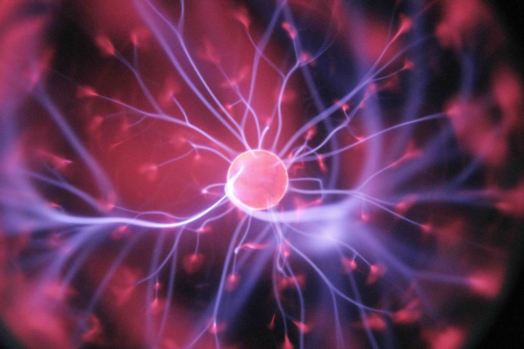
Header image by Hal Gatewood via Unsplash
