This post is also available in .
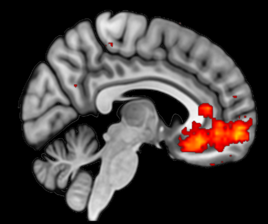
You have probably seen them before: those beautiful brain images with coloured ‘blobs’ that neuroscientists use to show brain activity. Such images are made with functional MRI (fMRI; Magnetic Resonance Imaging). We use this technique a lot at the Donders Institute. But why do we use an MRI scanner for this, and how does it actually work?
Measuring which brain areas are active
At the Donders Institute, we study how cognition and behaviour (e.g. perception, memory, or decision making) work in the brain. We often do this by letting test subjects carry out a task and see whether certain events (e.g. perceiving a stimulus, or making a decision) are associated with more brain activity in a particular area. In this way, we can deduce whether this brain area plays a particular role in the behaviour we are trying to understand.
An indirect measurement of brain activity: oxygen in the blood
Although we are interested in brain activity, the MRI scanner actually measures oxygen in the blood. However, these two are closely related.
As you may know, brain cells (neurons) communicate with each other using electrical and chemical signals. To send these signals, neurons need energy and oxygen and this must be supplied by blood. When a group of neurons becomes active, the body responds by sending lots of oxygen-rich blood to that brain area. In fact, the body supplies so much oxygen-rich blood that there is a net increase in oxygen-rich blood. This relationship between neuronal activity and oxygenated blood is called the blood oxygenation level-dependent (BOLD) response.
This BOLD response is what we measure with fMRI: it is therefore an indirect measurement of brain activity1.
Your brain in a big magnet
But how can we measure this increase in oxygenated blood? For this purpose, we use the magnetic properties of oxygenated and deoxygenated blood. Whereas oxygen-poor blood is very sensitive to magnetic fields, oxygen-rich blood is much less sensitive. We can measure this difference when we put people in a large electromagnet: this is the round tube of the MRI scanner in which you lie. In this way, we can distinguish between brain areas with more or less oxygen-rich blood, and thus deduce where there is more or less brain activity.
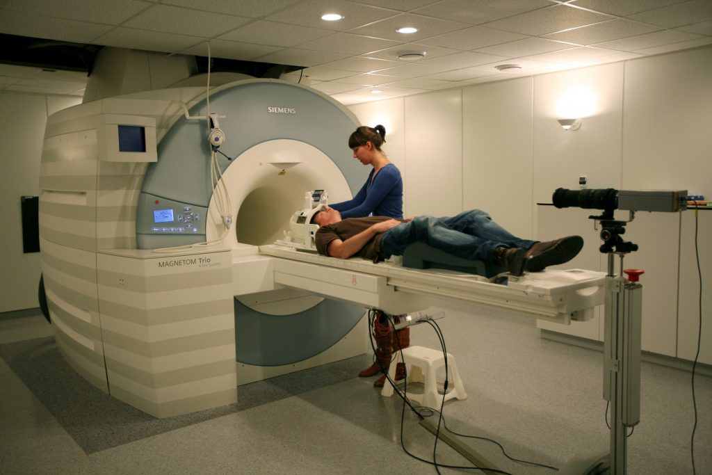
Hopefully you now understand why and how we measure brain activity with the MRI scanner. Would you like to experience this in real life? Then sign up for an MRI study!
1Oxygen in the blood is not only influenced by brain activity, but also by other physiological processes. That is why researchers often include all kinds of other measurements in their analyses, such as heart rate and respiration.
Credits
Author: Felix Klaassen
Buddy: Marlijn ter Bekke
Editor: Brittany van Beek
Translation: Ellen Lommerse
Editor translation: Christienne Damatac
Featured image constructed using Neurosynth
Curious to see what the MRI lab of the Donders looks like? Then watch this video:
1 thought on “How do we measure brain activity with an MRI scanner?”