This post is also available in Dutch .
Can fetuses participate in research studies? They can now, thanks to modern neuroimaging techniques and the field is rapidly growing. New engineering solutions will help to further develop our knowledge.
Many of us take what we know for granted, but our memories, habits, and behaviors are built up over many years of learning. The time we spend in the womb as a baby is a special period when the brain develops and undergoes major changes that will affect us for our entire lives. Observing the brain and behaviour of fetuses in the womb can answer many interesting questions such as: “Do fetuses like Mozart?” or, “When does intelligence start?”

But are these studies safe? Yes! By combining ultrasound and magnetic field scanners we can look into the fetal brain without disturbing them. Let’s start with a bit of history…
A set of revolutionary experiments in the early 2000 paved the way to a better understanding of fetus hearing and vision in the womb, but the main challenge is guessing where the fetus is! The development of better techniques opens a window to the study of consciousness during pregnancy. But, how exactly can we do it?
A window onto fetal thoughts
Did you know that fetuses can already hear sound during the 22nd week (which is around 5 months)? And did you know that fetuses can see? It must be quite dark in there!
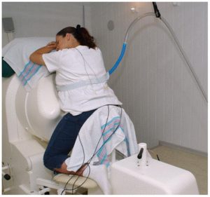
In our study we presented sounds and light flashes through the mother’s belly and measured the brain responses of fetuses to those stimuli. During the study, we also took care to estimate the position of the head, because we cannot see it directly (the womb is not transparent). We did this by combining the ultrasound images with magnetoencephalography, a scanning technique that measures magnetic fields coming from the womb. What we saw is that indeed the fetal brain responds to sounds and flashes, but only if we are able to localise the head accurately!
A complicated challenge
When researchers study the adult brain it is easy to get the participant to follow your instructions because subjects are intelligent and can interact with us.

Figure 2: A fetal magnetoencephalograpy scanner. Sound and light are delivered on the belly via a loudspeaker (blue tube) and light emitting diodes (LEDs). Picture courtesy of Cristiano Micheli (license)
We often try to get our participants to lie as still as possible to get the best test results, however it is impossible to ask a fetus to stop moving for a minute! So, simple as it sounds, how to manage movements during fetal recording is one of the biggest challenges for researchers at the moment. Of course, we are trying our best to solve this problem, and this challenge is the object of a new study direction that you can follow here. Stay tuned!
Written by Cristiano Micheli, edited by Monica Wagner, translated by Felix Klaassen
