This post is also available in Dutch.
Functional MRI (fMRI) has long been a core method to study brain function. Conventional fMRI, however, captures only a “zoomed out picture.” A more high-resolution version, laminar fMRI, lets scientists closely examine the different layers of the brain. But how does this work?
Beyond the ‘Blobs’: From Conventional to Laminar fMRI
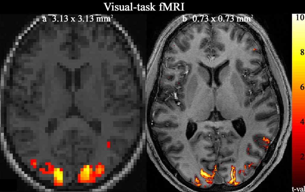
Conventional fMRI is a common neuroimaging technique that measures blood oxygenation level-dependent (BOLD) signal to reveal which brain regions respond to a task. In these scans, the brain is divided into small, 3D cubes called ‘voxels’ so researchers can see which of these voxels are active. For conventional fMRI, these voxels are often around 2mm3 which creates the characteristic fMRI “blobs”, the blurry shapes that you can see in left image below.
With voxels this size, researchers often capture signals across the entire thickness of the cortex. The cortex is the outer layer of the brain that controls thinking, sensing, and voluntary movements and is about 1-4.5 mm thick. However, this cortex is not uniform; it has six unique layers, each with distinct functions and connections. Laminar fMRI allows us to zoom in on these individual layers, revealing a new level of detail.
How does laminar fMRI work?
To reach the precision required to study cortical layers, scientists need very powerful MRI scanners. In MRI, a Tesla (T) is the unit used to measure the strength of the magnetic field. While traditional fMRI often uses 3T scanners, laminar fMRI typically requires 7T or stronger scanners. These stronger scanners allow for ‘sub-millimeter’ resolution with voxels smaller than 1mm³. This means laminar fMRI captures the unique activity in these smaller voxels and we can begin to “see” which layers are active during specific cognitive processes.

Conventional voxel size (left) versus submillimeter voxel size (right) during a visual task. Image by Yun and collegues (2023)
How Information Flows Across Layers
Why go to such lengths? Laminar fMRI can show how information moves across cortical layers, providing new insights into how the brain manages complex functions. For example, “feedforward” signals, which carry new sensory information, usually activate the superficial and middle layers of the cortex. “Feedback” signals, which adjust activity based on context or memory, tend to activate deeper layers. Different layers also connect to distinct brain regions, with deep layers linking the cortex to subcortical structures (like the thalamus) and middle layers focusing on connecting one area of the brain with another.
Until recently, most laminar fMRI studies focused on areas like the visual and motor cortex because they are easier to activate with simple tasks, such as finger-tapping to image the layers of the motor cortex. Nowadays, researchers can also image other areas, such as the prefrontal cortex. This makes it possible to study complex processes like perception, consciousness, hallucinations or working memory. For example, recent findings show that different prefrontal cortex layers respond uniquely during memory tasks—an exciting step forward.
The future of laminar fMRI
But laminar fMRI isn’t without challenges. It requires powerful, high-field scanners that produce noisier data, often necessitating longer scans or smaller target areas for better signal quality. Also, current technology can’t directly map all six layers, so signals are usually grouped into deep, middle, and superficial layers.
As hardware and software continue to improve, the potential for laminar fMRI grows. Soon, the strongest MRI scanner in the world with the strength of 14Twill be available at the Donders Institute, which may bring even deeper insights into how brain structure supports complex functions. Laminar fMRI could eventually give us a much more detailed understanding of the human brain at work.
Credits
Author: Helena Olraun
Buddy: Siddharth Chaturvedi
Editor: Vivek Sharma
Translation: Dirk-Jan Melssen
Editor translation: Lucas Geelen
