This post is also available in Dutch .
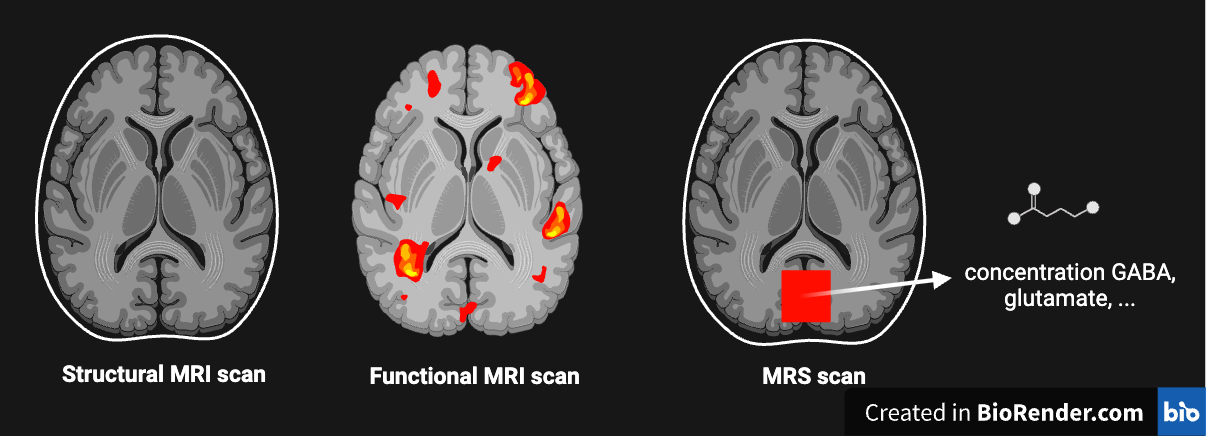
You may be familiar with the images of the brain that can be made with an MRI scanner: for instance, they can be used by a medical doctor to determine whether there is a tumour in the brain. Furthermore, a researcher can study brain activity using functional MRI. Medical doctors and researchers can now also use the MRI scanner to determine the concentration of certain molecules in the brain. This technique is called Magnetic Resonance Spectroscopy (MRS). But why are these molecules interesting to begin with?

The balance between excitation and inhibition
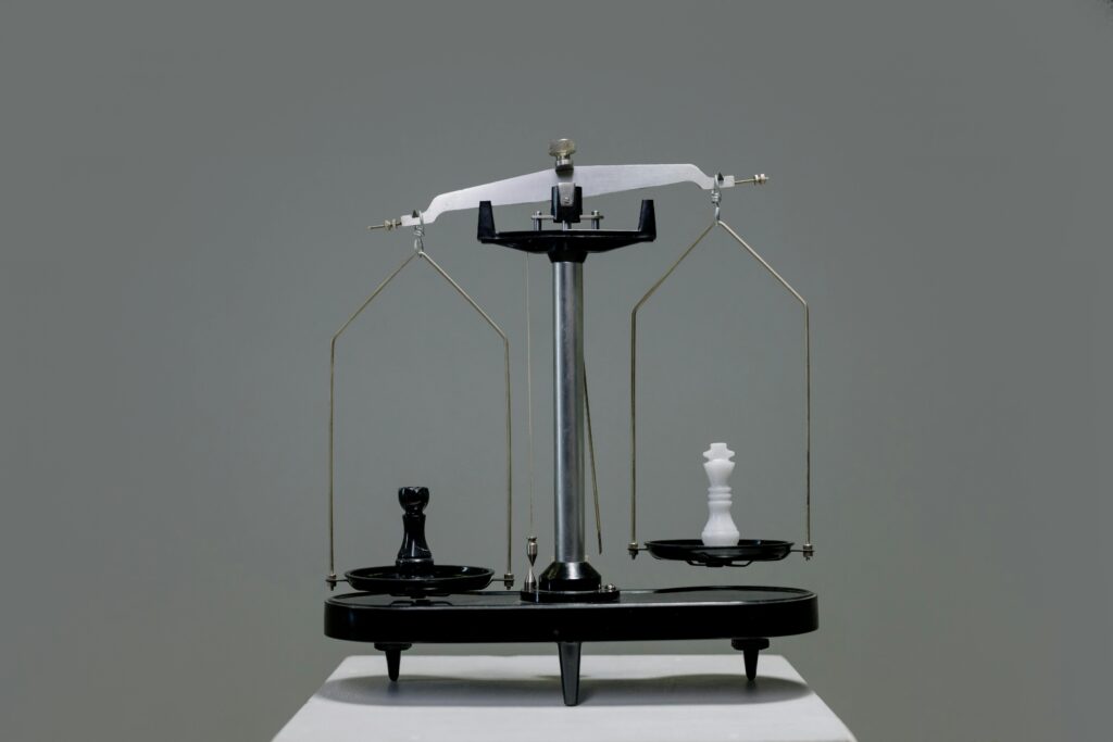
Our brain cells communicate with each other using signal molecules, also known as neurotransmitters. We can measure some of these neurotransmitters with MRS, including the molecule Gamma-Aminobutyric Acid (GABA). This substance plays an important role in brain functioning: GABA reduces activity in other brain cells, which is called inhibition. This is in contrast to most other neurotransmitters, which stimulate other brain cells. This excitation can happen through, for example, the substance glutamate. The balance between excitatory and inhibitory signals is crucial for healthy brain function. Without inhibition to keep it in check, excitation can lead to uncontrolled activity in the brain, resulting for instance in an epileptic seizure. With MRS we measure the substances that regulate this balance.
Fingerprints of signal molecules with MRS
The principle of MRS is based on the fact that every molecule, including GABA, exhibits slightly different behaviour in the magnetic field of the MRI scanner. This makes it possible to measure ‘fingerprints’ of all the molecules in a brain area. What makes MRS difficult compared to a ‘normal’ MRI scan is that interesting signalling molecules, such as GABA, are present in the brain only in relatively low concentrations and only give off little measurable signal. For this reason, neurotransmitters with even lower concentrations, such as dopamine or serotonin, cannot be properly studied (yet) with MRS. To still receive enough signal, MRS measures in an area about 1000x larger than a ‘normal’ MRI scan! Furthermore, measurements are taken over longer periods to create a single MRS scan.
The future of MRS
Thanks to the development of MRI scanners with stronger magnets (see also the plans to build the world’s strongest MRI scanner in Nijmegen!), it has become easier to study GABA and glutamate, because a stronger magnet is better for picking up their weak signals. Thanks to these stronger magnets and improved techniques, measurements take less time. As a result, researchers can now measure changes in glutamate and GABA over time, studying what happens to glutamate when a study participant looks at a blinking pattern, or recalls a memory. It is also now possible to measure glutamate simultaneously across multiple brain regions, with a much-improved resolution.
With these ever-improving techniques, we gain more and more insight into the role that the balance between neurotransmitters plays in our brain’s functioning. For example, recent research shows that the balance between excitation and inhibition plays a role in learning and decision-making. Evidence is also emerging that an imbalance between neurotransmitters may underlie conditions such as Post-Traumatic Stress Disorder (PTSD). But of course, the more we discover, the more questions we have!
Credits
Author: Renée Koolschijn
Buddy: Helena Olraun
Editor: Lucas Geelen
Translation: Renée Koolschijn
Editor translation: Helena Olraun
Image: cottonbro studio via pexels
